Glaucoma. Diabetes. Macular Degeneration. Cataract.
|
Glaucoma
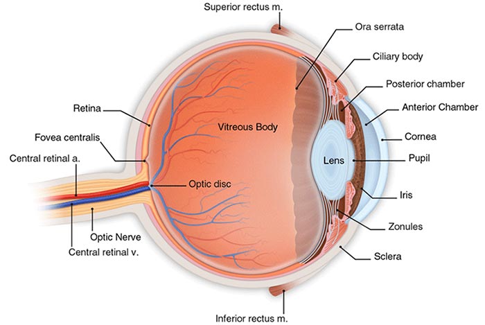
What is Glaucoma? Glaucoma is a group of diseases that can damage the optic nerve in the eye. If left untreated, glaucoma can cause permanent vision loss or blindness. What causes Glaucoma? Clear fluid flows in and out of a small space at the front of the eye called the anterior chamber. This fluid bathes and nourishes nearby tissues. If this fluid drains too slowly, pressure builds up and damages the optic nerve. Though this buildup may lead to increase in eye pressure, the effect of pressure on the optic nerves differs from person to person. Some people may get optic nerve damage at low pressure levels while others tolerate higher pressure levels. Who is most likely to get it? Millions of people have glaucoma. Anyone can get it, but some people are at a higher risk: African Americans over the age of 40, Everyone over the age of 60, especially Mexican Americas, and people with a family history of glaucoma. What are the symptoms? At first, there are no symptoms. Vision stays normal and there is no pain. But as the disease gets worse, side vision may begin to fail. Objects straight ahead may be clear, but objects to the side might be missed. If left untreated, the field of vision narrows and objects in the front can no longer be seen. How is it detected? Glaucoma is found most often during a dilated eye exam. The doctor will put drops in the eyes to enlarge the pupils. This allows the doctor to see more of the inside of the eye to check for signs of damage. How can it be treated? Primary open-angle glaucoma cannot be cured, however it can usually be controlled. There are several medications in the form or eye drops of pills that can be used. For extreme cases, a consultation with an Ophthalmologist and a surgical procedure can help. (National Eye Institute) Macular Degeneration
What is Age-related Macular Degeneration? AMD is a common eye disease associated with aging that gradually destroys sharp, central vision. The retina is a paper-thin tissues that lines the back of the eye and sends visual signals to the brain. In the middle of the retina is a tiny area called the macula. The macula is made up of millions of light-sensing cells that help to produce central vision. How does Macular Degeneration damage vision? Dry Age-related Macular Degeneration affects 90% of AMD patients. Studies suggest that an area of the retina becomes diseased, leading to the slow breakdown of the light-sensing cells in the macula. Wet Age-related Macular Degeneration affects 10% of AMD patients. As DRY AMD worsens, new blood vessels may begin to grow and cause "wet" AMD. These new blood vessels will often leak blood and fluid under the macula. The causes rapid damage to the macula and can lead to loss of vision in a short period of time. Who is most likely to get AMD? The greatest risk factor is age. Studies show that people over the age of 60 are at greater risk than other age groups. Other factors include, Gender (women tend to be higher risk), Race (Caucasian's are more likely to lose vision), Smoking, and Family History. What are the symptoms? AMD causes no pain. The most common early sign of Dry AMD is blurred vision. The classic early symptom of Wet AMD is that straight lines appear crooked. How is it detected? A dilated retinal exam where the doctor places drops in the eyes to enlarge the pupil, allowing the doctor to see more of the retina and look for signs of macular degeneration. They will also use what is called an Amsler Grid to test your central vision. How can it be treated? No treatment exists for Dry AMD, some vitamins and minerals may help slow the progress of the disease. Some cases of Wet AMD can be treated with surgery. (National Eye Institute) To find out more information about these conditions visit the National Eye Institue Website
|
Diabetic Retinopathy
What is the retina? The retina is a light sensitive tissue at the back of the eye. When light enters the eye, the retina changes the light into nerve signals. The retina then sends these signals along the optic nerve to the brain. Without a retina, the eye cannot communicate with the brain, making vision impossible. How does diabetic retinopathy damage the retina? Diabetic retinopathy occurs when diabetes damages the tiny blood vessels in the retina. Some people may develop Macular Edema, which occurs when the damaged blood vessels leak fluid and lipids onto the macula. The fluid makes the macula swell, blurring vision. As the disease progresses, fragile, new blood vessels grow along the retina and in the clear, gel-like vitreous that fills the inside of the eye. These new blood vessels can bleed, cloud vision, and destroy the retina. Who is at risk for this disease? All people with diabetes are at risk, including Type I, Type II, and Gestational Diabetes. What are its symptoms? There are no early warning signs. At some point, though, you may have macular edema. It blurs vision. In some cases, you vision will get better or worse during the day. How is it detected? During a comprehensive eye exam, the doctor will test your visual acuity, how well you see at various distances. They will perform a dilated retinal exam, placing drops in the eyes to enlarge the pupil, allowing the doctor to see more of the retina and look for signs of diabetic retinopathy. During that dilated retinal exam, the doctor will also perform Opthalmoscopy. Using a device with a special magnifying glass, the doctor gains a wide view of the retina. How is it treated? There are two types of treatment. One is Laser Surgery performed by an Ophthalmologist. The other is Vitrectomy, a surgery that will remove the blood filled vitreous and replace it with a salt solution. (National Eye Institute) Cataract
What is a cataract? A cataract is a clouding of the eye's lens that causes loss of vision. What causes it? The lens lies behind the iris and the pupil. It works much like a camera lens. It focuses light onto the retina at the back of the eye, where an image is recorded. The lens also adjusts the eye's focus, letting us see things clearly both up close and far away. The lens is made of mostly water and protein. The protein is arranges in a precise way that keeps the lens clear and lets light pass though it. As we age, some of the protein may clump together and start to cloud a small area of the lens. This is a cataract. Over time, the cataract may grow larger and cloud more of the lens, making it harder to see. When are you most likely to have a cataract? People can have a cataract in their 40's and 50's but during middle age, cataracts are small and do not affect vision. It is after age 60 that most cataracts steal vision. What are it's symptoms? You may notice that your vision is blurred a little, like looking through a cloudy piece of glass. Light from the sun or a lamp may seem too bright, causing a glare or you may notice when you drive at night that the oncoming headlights cause more glare than before. Colors may not appear as bright to you as they once did and as the cataract gets bigger, you will find it harder to read and do other normal tasks. How is a cataract detected? The only way to know for sure is to have an eye examination. How is a cataract treated? Surgery is the only way to treat cataract. You doctor will remove your clouded lens and replace it with a clear, plastic lens. Cataract surgery is very successful in restoring vision. It is one of the most common surgeries performed in the United States, with over 1.5 million cataract surgeries done each year. (National Eye Institute) |

